Knee Fracture
Knee is composed of mainly 3 bone femur, patella and tibia. Hence, knee fractures are mainly related to these three bones.
Distal Femur Fracture
Although less frequent than fractures around the hip, distal femur fractures are relatively common injuries and present considerable challenges in management.
Internal fixation is recommended for most displaced distal femoral fractures in adults.
The goals of treatment are:
- Anatomic reduction of the articular surface
- Restoration of limb alignment, length, and rotation
- Stable fixation that allows for early mobilization
Mechanisms of injury for distal femur fractures
Distal femur fractures can occur in the young as well as the elderly population. In young patients significantly high energy trauma is required to cause a fracture whereas in an elderly population a low energy trauma can also cause a fracture.
Signs and symptoms of distal femur fractures:
- Swelling
- Pain
- Deformity
- Altered leg alignment with shortening
- Internal bleeding may cause the skin around the knee to become black & blue
- A careful neurovascular examination must be performed and documented including the presence or absence of distal pulses and sensorimotor assessment
Imaging and other diagnostic studies for distal femur fractures:
- Anteroposterior (ap) and lateral radiograph of the knee and femur
- X-rays of the pelvis, ipsilateral hip, and femoral shaft are necessary to rule out associated injuries
- CT Scans
Classification of distal femur fractures:
Essentially all classifications distinguish among
- Extra-articular
- Intra-articular
- Isolated condylar lesions
Fractures are further subdivided according to the degree and direction of displacement, amount of comminution, and involvement of the joint surfaces.
Factors which play a dynamic role in management are:
- Amount of fracture displacement
- Degree of comminution
- Extent of soft tissue injury
- Associated neurovascular injuries
- Magnitude of joint involvement
- Degree of osteoporosis
- Presence of multiple trauma
- Complex ipsilateral injuries (i.e., Patella or plateau fractures)
Distal femur fracture treatment options:
Nonoperative treatment of distal femur fractures
Indication:
- Reliable patients with a nondisplaced fracture
- Nonambulatory patients (e.g., Paraplegia)
- Patients with significant underlying medical diseases (e.g., Severe Cardiopulmonary Risk) or Imminent Death
- Infected fractures or severely contaminated open fractures (e.g., Type IIIB)
Treatment:
Closed reduction with skeletal traction with or without subsequent cast bracing. This method requires confinement to bed, is time consuming and morbid.
Operative treatment of distal femur fractures:
Indication:
All displaced distal femur fractures in physiologically stable adults
Types of Procedures:
- Osteosynthesis of distal femur fractures with plates and screws (open reduction and internal fixation)
- Retrograde femoral nailing of distal femur fractures
- Intramedullary nailing of distal femur fractures with flexible and semirigid nails
- External fixation of distal femur fractures
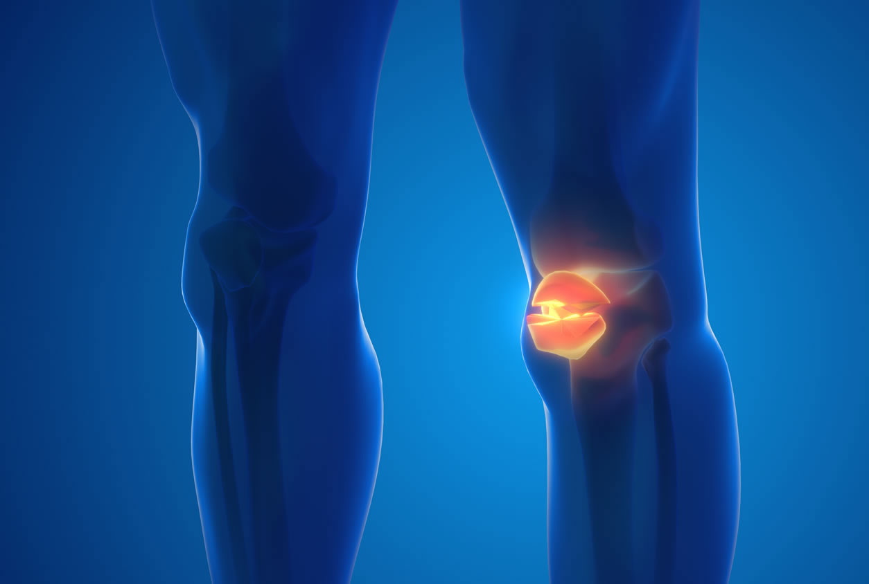
Patella Fracture
Patella is the largest sesamoid bone in the body. A sesamoid bone is one that grows within a tendon. The clinical picture of a patellar fracture is determined by a combination of definite and equivocal signs
Mechanism of injury
Direct Trauma: This is due to dashboard injuries and due to direct fall over the patella. They usually cause comminuted fractures, and are the common causes.
Indirect Trauma (Quadriceps Contraction): Sudden forceful contraction of the quadriceps as in sports person and athletes can cause patellar fractures. Here the fracture is usually transverse and sometimes avulsion fractures of the proximal or distal poles may be seen.
Classification
Undisplaced:
- Transverse fracture — These account for nearly 50-80 percent of cases. About 80 percent occur in the middle-third.
- Stellate Fracture
- Vertical Fracture
Displaced: If displacement is > 3 mm and if articular incongruity > 2 mm:
- Transverse — Involving upper or lower poles (50-85%).
- Oblique Fracture – Vertical Fracture (12-27%).
- Comminuted Fracture (30-35%)
- Polar — Could be Proximal or Distal
- Osteochondral Fractures
Clinical Features:
- The patient gives history of trauma following which there is pain and swelling at the knee joint.
- The patient is unable to extend the knee and both the active and passive movements are restricted.
- On examination, there could be a palpable gap, tenderness, signs of effusion and a positive patellar tap
Investigations
- Radiograph of the knee joint consists of ap view, lateral view, intercondylar notch view and skyline or axial view to rule out undisplaced vertical fracture.
- CT Scan, bone scan and tomography are other useful investigations
Management
Undisplaced Fracture:
Nonoperative treatment will produce good results in undisplaced fracture and if displacement is less than 1-2 mm and in intact extensor mechanism and minimal articular step-off (< 1-2 mm) and the methods include compression bandage, ice applications, aspiration of hemarthrosis, cylindrical cast in extension or long leg cast for 4-6 weeks. Functional cast brace is also effective. The patient is advised early weight bearing and quadriceps exercises.
- Lateral Retinacular Release: By open or arthroscopic technique, if incongruity and lateral tilting of the patella is seen on axial radiographs
- Osteotomies for bony malalignment, such as femur anteversion, tibial external torsion and genu valgum.
Displaced Fracture:
In this variety, surgery is the treatment of choice. Surgery is performed as early as possible preferably within 7 days.
Surgical Methods
Open reduction and internal fixation:
- This is indicated in transverse fractures of the patella. Internal fixation is done either by the circumferential wiring or by tension band wiring.
- The other methods are pyrford technique of circumferential wiring and a second tension band wiring through the tendon provide better fixation.
- Lotke Longitudinal Anterior Band (LAB) wiring is another method with good results.
Patellectomy:
- This could be either partial (for smaller distal or proximal pole fracture) or complete (for communited fractures). Disadvantages of patellectomy:
- Strength of quadriceps returns slowly although knee motion is regained quite fast.
- Obvious atrophy of the quadriceps muscle persists for months and often permanently.
- Protection of the knee by the patella is lost.
- Pathological ossification may develop where the patella is excised.
Proximal Tibial Fractures:
Proximal Tibia consists of the medial and lateral condyles along with the upper tibial articular surface and includes the proximal 10-12 cm of the tibia. These fractures are frequently intra - articular and usually unite well considering the cancellous nature of the bone.
Mechanism of injury:
It is due to valgus or varus force with axial loading.
Causes
- Due to auto-pedestrian injuries (bumper injuries).
- Due to fall from heights.
- Miscellaneous causes (football or soccer injuries).
Types
- Articular extension
- Extra-articular
Schatazker's classification:
This is widely followed in north america and has six types:
- Type i: Split fracture of lateral condyle.
- Type ii: Displaced lateral condyle fracture.
- Type iii: Isolated lateral condyle depression.
- Type iv: Medial condylar fracture.
- Type v: Bicondylar fractures.
- Type vi: Bicondylar fracture with diaphyseal metaphyseal extension.
Clinical Features
- Pain
- Swelling
- Deformity
- Haemarthrosis
- Decreased movements of the knee
- Instability in valgus or varus
- Compartment syndrome of the leg
- Disturbed peripheral vascular and nerve functions of the leg
Investigations:
- The routine ap and lateral radiographs of the knee help to demonstrate majority of tibial condyle fractures. Oblique view may be required to localize the fractures.
- To study the depth of depression, ct scan is excellent.
- To know the knee ligament injuries, valgus or varus stress films are required.
- Aspiration may reveal blood or fat. If fat is present, it indicates an intra-articular fracture.
- Angiography if pulses are feeble or absent.
Management
Aim
- To produce a knee that extends fully and flexes to at least 120°.
- Restoration of normal articular surface and ligament repair are both important in preventing late instability.
Conservative Treatment
Indicated for plateau fractures with < 4 mm depression or displacement.
Undisplaced Fracture:
Above knee, pop cast with 5° flexion or cast bracing is used.
Displaced Fracture:
Closed reduction, with or without skeletal traction and a long leg cast is used.
In Depressed Fractures:
- For less than 8 mm depression, above knee cast.
- For depression of more than 8 mm with a large split fragment, skeletal traction is applied.
- For more than 8 mm with smaller split fragment, orif is done with bone grafting after elevation of the depression.
Surgery
- In displaced condylar fractures, bicondylar fractures, split fractures, closed reduction is not useful.
- Open reduction and internal fixation with cancellous screws, single or dual buttress plating are the time tested methods.
- External fixation with circular or semi-circular frames are also another useful options.
- Skeletal traction is useful in grossly comminuted fractures.
To know more kindly contact the best orthopaedic surgeon Dr. Rahul Modi for further queries.
This surgery is frequently performed by the best orthopaedic surgeon Dr. Rahul Modi for treating Knee Fracture.